
 Cardiovascular Physiology Concepts, 3rd edition textbook, Published by Wolters Kluwer (2021)
 Normal and Abnormal Blood Pressure, published by Richard E. Klabunde (2013)
Action Potentials
Numerous cells in the body can undergo a transient depolarization and repolarization. This can be triggered by external mechanisms (e.g., motor nerve stimulation of skeletal muscle or cell-to-cell depolarization in the heart) or by intracellular, spontaneous mechanisms (e.g., cardiac pacemaker cells).
There are three general types of cardiac action potentials that are distinguished, in part, by the presence or absence of spontaneous pacemaker activity and by how rapidly they depolarize.
- Non-pacemaker action potentials, also called fast response action potentials because of their rapid depolarization, are characteristic of atrial and ventricular myocytes.
- Pacemaker cells generate spontaneous action potentials that are also termed slow response action potentials because of their slower rate of depolarization. These are found in the sinoatrial and atrioventricular nodes of the heart.
- Specialized conducting cells found within the His-Purkinje system of the ventricles are a third type of action potential that are fast response type action potentials; however, in contrast to normal atrial and ventricular myocytes (fast response, non-pacemaker cells), these His-Purkinje cells exhibit spontaneous depolarization.
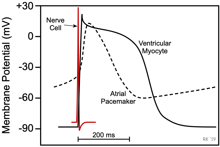 Cardiac action potentials differ considerably from action potentials found in neural and skeletal muscle cells. One major difference is in the duration of the action potentials. In a typical nerve, the action potential duration is about 1 ms. In skeletal muscle cells, the action potential duration is approximately 2-5 ms. In contrast, the duration of cardiac action potentials ranges from 200 to 400 ms. Another difference between cardiac and nerve and muscle action potentials is the role of calcium ions in depolarization. In nerve and muscle cells, the depolarization phase of the action potential is caused by an opening of fast sodium channels. This also occurs in non-pacemaker cardiac cells; however, in cardiac pacemaker cells, calcium ions are involved in the initial depolarization phase of the action potential. In non-pacemaker cells, calcium influx prolongs the duration of the action potential and produces a characteristic plateau phase.
Revised 11/01/2023