
 Cardiovascular Physiology Concepts, 3rd edition textbook, Published by Wolters Kluwer (2021)
 Normal and Abnormal Blood Pressure, published by Richard E. Klabunde (2013)
Electrocardiogram Chest Leads (Unipolar)
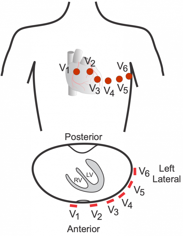 Besides the three standard limb leads and the three augmented limb leads that view the electrical activity of the heart from the frontal plane, there are six precordial, unipolar chest leads. This configuration places six positive electrodes on the surface of the chest over different regions of the heart to record electrical activity in a plane perpendicular to the frontal plane (see figure). These six leads are named V1 – V6. The rules of interpretation are the same as for the limb leads. For example, a wave of depolarization traveling toward a particular electrode on the chest surface will elicit a positive deflection.
The chest leads overlie the following ventricular regions:
| Leads | Ventricular Region |
| V1-V2 | anteroseptal |
| V3-V4 | anteroapical |
| V5-V6 | anterolateral |
This placement of chest leads produces the following normal ECG tracings:
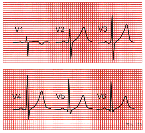 Because initial ventricular depolarization is from left to right across the septum, there is an initial R-wave in V1 followed by an S-wave as the anterior and lateral walls of the left ventricle depolarize. Leads V5 and V6 have a large net positive QRS because these leads overlie the anterolateral wall of the left ventricle, which has a large muscle mass undergoing depolarization. Tracings from leads V5 and V6 are almost opposite in polarity from V1 because they are viewing opposite sides of the heart. Leads V2-V4 are intermediate owing to their electrode placement.
In summary, the chest leads provide a different view of the electrical activity within the heart. Therefore, the waveform recorded is different for each lead compared to the limb leads. To understand how cardiac electrical currents actually generate and ECG tracing and why the different leads display that electrical activity differently, it is necessary to understand volume conductor principles and vectors.
See also:
Revised 02/15/2024