
 Cardiovascular Physiology Concepts, 3rd edition textbook, Published by Wolters Kluwer (2021)
 Normal and Abnormal Blood Pressure, published by Richard E. Klabunde (2013)
Volume Conductor Principles and ECG Rules of Interpretation
The electrocardiogram uses electrodes on the surface of the body to measure the electrical activity of the heart. It is possible to place electrodes on the body surface to measure cardiac electrical activity because the body acts as a conductor of the electrical currents generated by the heart. What do these electrodes actually measure?
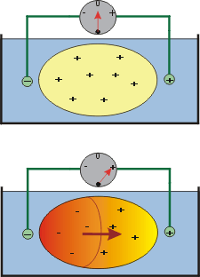 If a piece of living ventricular muscle is placed in a bath containing a salt solution to conduct electrical currents, and electrodes are placed in the bath on either side of the muscle, no potential difference would be recorded between the two electrodes when the muscle is in its polarized, resting state (top panel of figure at right). This is because the outside of the cells is positive relative to the inside because the resting membrane potential is about -90 mV; therefore, no currents will flow along the surface of the muscle and through the bath. If the left side of the muscle is stimulated electrically to induce self-propagating action potentials, a wave of depolarization would sweep across the muscle from left-to-right (lower panel). Midway through this depolarization process, cells on the left (depolarized cells) would be negative on the outside relative to the inside, while non-depolarized cells on the right of the muscle would still be polarized (positive on the outside). There would now exist a potential difference between the positive and negative electrodes. By convention, a wave of depolarization heading toward the positive electrode is recorded as a positive voltage (upward deflection in the recording). After the wave of depolarization sweeps across the entire muscle mass, all the cells on the outside are negative, and once again, no potential difference would exist between the two electrodes.
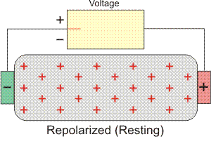 The entire process of depolarization and repolarization is depicted in the animated model to the right, which is representative of the electrical events that occur in the atria. In the resting, polarized state, no potential difference is measured between the positive and negative electrodes (i.e., isoelectric – flat red line). When the left side of the tissue becomes depolarized (representing firing of the SA node), a wave of depolarization begins to spread across the atria. During this time, some muscle mass temporarily remains positive on the outside (polarized) and while some is negative (depolarized); thus, there is a separation of charges which causes a potential difference between the two electrodes. Because the wave of depolarization is moving toward the positive electrode, by convention, a positive voltage (upward deflection) is recorded. The voltage reaches its maximal positive value when half the tissue is depolarized. Once the entire atrial mass is depolarized (all cells negative on the outside), there is no longer a potential difference and the voltage is zero just as it was in the polarized state. When repolarization occurs, starting first with the left side (SA nodal region) then moving across the atria, there will once again be both positive and negative charges on the surface of the atria, but this time, the negative charges will be closest to the positive electrode. The wave of repolarization sweeping across the atria away from the negative electrode and toward the positive electrode causes, by convention, a negative voltage (downward deflection) to occur. Finally, when all the cells are repolarized, the measured voltage difference will once again be zero until another wave of depolarization occurs.
 A similar process occurs within the ventricles, with one major difference: repolarization normally occurs in a direction opposite to depolarization. In other words, the last cells in the ventricle to depolarize are the first to repolarize. This results in a positive recording as the ventricles repolarize as shown in the animated model to the right.
Several important observations and rules emerge from these volume conductor considerations:
- A wave of depolarization traveling toward a positive electrode produces a positive deflection in the ECG trace.
- A wave of depolarization traveling away from a positive electrode produces a negative deflection.
- A wave of repolarization traveling toward a positive electrode produces a negative deflection.
- A wave of repolarization traveling away from a positive electrode produces a positive deflection.
- A wave of depolarization or repolarization traveling perpendicular to an electrode axis produces no net deflection.
- The instantaneous amplitude of the measured potential depends on the direction of the mean electrical vector relative to the recording electrode lead axis.
- The voltage amplitude is directly related to the mass of tissue undergoing depolarization or repolarization.
The first four rules are derived from the volume conductor model described above. The fifth rule is also based on volume conductor principles and could be modeled by placing the positive and negative electrodes midway on the top and bottom surfaces of the tissue instead of on the ends. In this case, the positive electrode would first measure a positive voltage as the wave of depolarization transverse the tissue from the left edge to the midpoint (toward the electrode), and then the electrode would measure a negative voltage as the wave moved away from the electrode to the right edge. The net voltage recorded would be zero. The sixth rule considers that at any given time during depolarization in the atria or ventricles, there are many waves of depolarization traveling in different directions relative to the positive electrode. The recording by the electrode reflects the average, instantaneous direction and magnitude (i.e., mean electrical vector) for all the individual depolarization waves. The seventh rule simply states that the amplitude of the wave recorded by the ECG is directly related to the mass of the muscle undergoing depolarization or repolarization. For example, when the mass of the left ventricle is increased by hypertrophy, the voltage amplitude of the QRS complex, which represents ventricular depolarization, is increased in certain leads.
Revised 11/02/2023