
 Cardiovascular Physiology Concepts, 3rd edition textbook, Published by Wolters Kluwer (2021)
 Normal and Abnormal Blood Pressure, published by Richard E. Klabunde (2013)
Overdrive Suppression
Although the primary pacemaker site within the heart is the SA node, other cells have pacemaker activity (automaticity) or can become pacemakers under special conditions. These can be normal cells, such as those in the AV node and Purkinje fibers, or they can be other cells that display automaticity because hypoxic conditions have triggered pacemaker currents.
The SA node is normally the dominant, driving pacemaker because it has the highest intrinsic rate of spontaneous automaticity. For example, pacemaker sites within the ventricles typically have a rate of 30 – 40 depolarizations per minute, whereas cells within the AV node and bundle of His have an intrinsic rate of 40 – 50 depolarizations per minute. In contrast, the normal resting sinus rate is 60 – 100 depolarizations per minute, although it can be much higher under conditions of sympathetic activation.
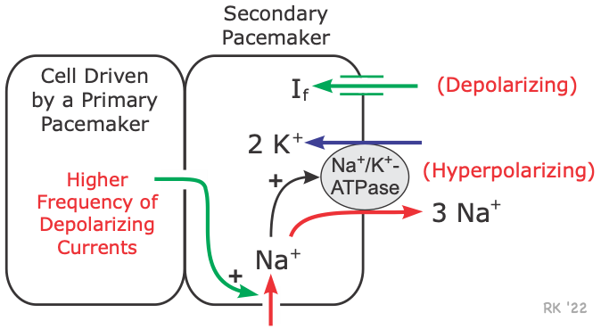 The higher frequency of SA nodal firing suppresses other pacemaker sites by a mechanism called overdrive suppression. If a latent pacemaker is being depolarized at a higher frequency than its intrinsic rate by an adjacent cell that is driven by the primary pacemaker, then the increased frequency of depolarizations leads to an increase in intracellular sodium ions. This occurs because more sodium ions enter the cell per unit time at a higher frequency. This increased sodium stimulates the Na+-K+-ATPase (increases its activity) to expel more sodium from the cell in exchange for potassium (see figure). Because this pump is electrogenic, increased pump activity increases hyperpolarizing currents generated by the pump. This drives the membrane potential more negative, thereby offsetting the depolarizing pacemaker currents (If) being carried into the cell. This effectively prevents the pacemaker currents from depolarizing the cell to its threshold potential and prevents the spontaneous generation of action potentials. If the cell ceases to be driven by the SA node (e.g., because of AV block), then the additional hyperpolarizing currents will be lost and spontaneous depolarization and action potential generation can occur.
Revised 11/02/2023