
 Cardiovascular Physiology Concepts, 3rd edition textbook, Published by Wolters Kluwer (2021)
 Normal and Abnormal Blood Pressure, published by Richard E. Klabunde (2013)
Cardiac Cycle - Atrial Contraction (Phase 1)
A-V Valves Open; Semilunar Valves Closed
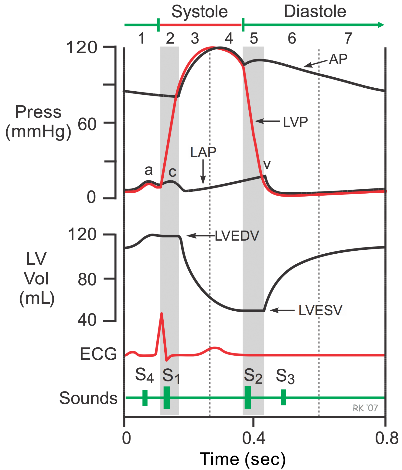 This is the first phase of the cardiac cycle. Electrical depolarization of the atria (corresponding to the P wave of the ECG) starts this phase of atrial muscle contraction. As the atria contract, the pressure within the atrial chambers increases, which forces more blood flow across the open atrioventricular (AV) valves, leading to a rapid flow of blood into the ventricles. Blood does not flow back into the vena cava because of inertial effects of the venous return and because the wave of contraction through the atria moves toward the AV valve, producing a "milking effect." Atrial contraction produces a small, transient increase in atrial and venous pressures called the a-wave. The x-descent follows the peak of the a-wave.
Atrial contraction normally accounts for about 10% of left ventricular filling when a person is at rest because most of ventricular filling occurs before atrial contraction as blood passively flows from the pulmonary veins, into the left atrium, then into the left ventricle through the open mitral valve.
At high heart rates, when there is less time for passive ventricular filling, atrial contraction may account for up to 40% of ventricular filling. This is sometimes referred to as the "atrial kick." The atrial contribution to ventricular filling varies inversely with the duration of ventricular diastole and directly with atrial contractility.
After the atrial contraction is complete, the atrial pressure falls, causing a pressure gradient reversal across the AV valves. This causes the valves to float upward (pre-position) before closure. The ventricular volumes are maximal, which is termed the end-diastolic volume (EDV). The left ventricular EDV (LVEDV), which is typically about 120 ml, represents the ventricular preload and is associated with end-diastolic pressures of 8-12 mmHg and 3-6 mmHg in the left and right ventricles, respectively.
Atrial contraction is sometimes associated with a fourth heart sound (S4), that may be caused by vibrations of the ventricular wall during atrial contraction. An S4 may be noticed when the ventricle compliance is reduced ("stiff" ventricle) as occurs in ventricular hypertrophy and in many older individuals.
Jump to other phases:
- Phase 2 - Isovolumetric Contraction
- Phase 3 - Rapid Ejection
- Phase 4 - Reduced Ejection
- Phase 5 - Isovolumetric Relaxation
- Phase 6 - Rapid Filling
- Phase 7 - Reduced Filling
Revised 11/04/2023