
 Cardiovascular Physiology Concepts, 3rd edition textbook, Published by Wolters Kluwer (2021)
 Normal and Abnormal Blood Pressure, published by Richard E. Klabunde (2013)
Microcirculation Structure and Function
The microcirculation comprises arterioles, capillaries, venules, and terminal lymphatic vessels.
Arterioles
- Small precapillary resistance vessels (10-200 μ) composed of an endothelium surrounded by one or more layers of smooth muscle cells
- Richly innervated by sympathetic adrenergic fibers and highly responsive to sympathetic vasoconstriction via both α1 and α2 postjunctional receptors.
- Represent the major site for regulating systemic vascular resistance.
- Rhythmical contraction and relaxation of arterioles sometimes occurs (i.e., spontaneous vasomotion).
- Primary function of arterioles within an organ is flow regulation, which determines oxygen delivery and the washout of metabolic by-products from the tissue.
- Regulate, in part, capillary hydrostatic pressure and therefore influence capillary fluid exchange.
Capillaries
- Small exchange vessels (6-10 μ) composed of highly attenuated (very thin) endothelial cells surrounded by basement membrane − no smooth muscle.
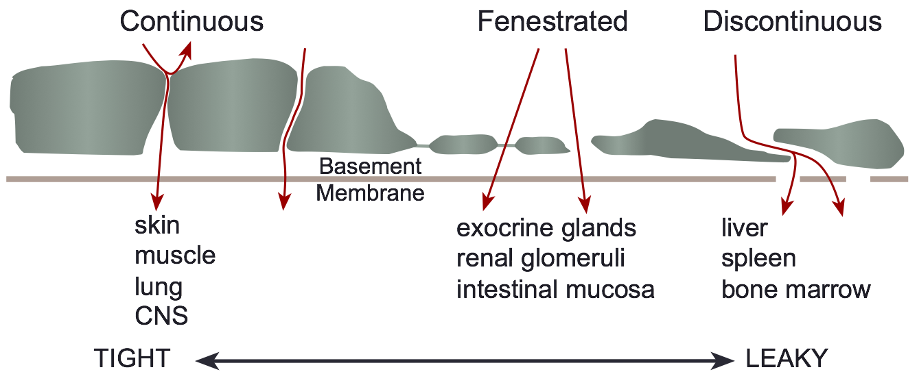 Three structural classifications:
- Continuous (found in muscle, skin, lung, central nervous system) − basement membrane is continuous and intercellular clefts (between adjacent endothelial cells) are tight (i.e., have tight junctions); these capillaries have the lowest permeability.
- Fenestrated (found in exocrine glands, renal glomeruli, intestinal mucosa) − perforations (fenestrae) in endothelium result in relatively high permeability.
- Discontinuous (found in liver, spleen, bone marrow) − large intercellular gaps and gaps in basement membrane result in extremely high permeability.
- Large surface area and relatively high permeability (especially at intercellular clefts) to fluid and macromolecules make capillaries the primary site of exchange for fluid, electrolytes, gases, and macromolecules.
- In some organs, the number of perfused capillaries can increase during high flow states (e.g., in contracting muscle) in response to arteriolar vasodilation and redistribution of precapillary pressures
Venules
- Small exchange vessels (10-200 μ) composed of endothelial cells surrounded by basement membrane (smallest postcapillary venules) and smooth muscle (larger venules).
- Fluid and macromolecular exchange occur at small postcapillary venular junctions.
- Sympathetic innervation of larger venules can alter venular tone, which plays a role in regulating capillary hydrostatic pressure.
Terminal Lymphatics
- Composed of endothelium with intercellular gaps surrounded by highly permeable basement membrane and are similar in size to venules − terminal lymphatics end as blind sacs.
- Larger lymphatics also have smooth muscle cells.
- Spontaneous and stretch-activated vasomotion is present, which serves to pump lymph out of the tissue.
- Sympathetic nerves can modulate vasomotion and cause contraction.
- One-way valves direct lymph away from the tissue and eventually back into the systemic circulation via the thoracic duct and subclavian veins (2-4 liters/day returned).
Revised 11/06/2023